
★企業と人材のマッチングサービスを準備中です。アンケートのご協力をお願いいたします!↓

最終更新日:2020/03/10
[論文] “Searching for prostate cancer by fully automated magnetic resonance imaging classification: deep learning versus non-deep learning”. Scientific Reports, 7, 15415 (2017).
[DOI: 10.1038/s41598-017-15720-y]
要点紹介
✔️前立腺癌の識別は非常に重要であるが、その検出精度を高めることは長年の課題であった
✔️前立腺癌患者と前立腺良性症状患者を識別するために、ディープラーニングを用いた
✔️ディープラーニングを用いた自動検出は、ディープラーニングを使わない場合よりも高い精度で前立腺癌患者を識別できることが示された
概説
前立腺癌(Prostate Cancer;PCa)は古代エジプトのプトレマイオスのミイラの画像に記録されて以来の主な死因である。
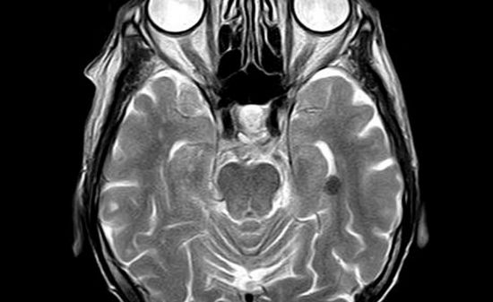
PCa検出は個別化医療にとって極めて重要であり、MRIスキャンにおいては非常に多様である。172人の患者に対して、2,602の前立腺の形態学的画像(アキシャル2D T2強調画像)が得られた。
病理学的に確認されたPCa患者と前立腺炎や良性前立腺過形成(BPH)といった前立腺の良性症状(BC)患者を区別するために、深層畳み込みニューラルネットワーク(DCNN)を用いたディープラーニング、ならびに画像認識および分析の代表的な手法であるSIFT画像特徴やバッグオブワード(BoW)を用いたノンディープラーニングを用いた。
PCa患者の完全自動検出では、ディープラーニングはノンディープラーニングよりも受信者動作特性曲線(AUC)において統計的に高い領域にあった(P = 0.0007 < 0.001)。
AUCは、ディープラーニングで0.84(95%CI 0.78-0.89)、ノンディープラーニングで0.70(95%CI 0.63-0.77)であった。
我々の結果は、DCNNを用いたディープラーニングが、前立腺BC患者と完全自動PCa患者検出に関して、SIFT画像特徴やBoWモデルを用いたノンディープラーニングよりも優れていることを示唆している。
我々のディープラーニング法は、MRIやCT、他の臓器のPET検査などの画像診断に拡張可能である。

SIFT画像特徴およびBoWモデルによるノンディープラーニングと、深層畳み込みニューラルネットワーク(DCNN)によるディープラーニングとの区別を示している。
注)ROC曲線:受信機動作特性曲線 AUC:ROC曲線の下の領域 PCa:前立腺癌 前立腺BC:前立腺良性状態 BPH:良性前立腺肥大
AIDBの全記事が読み放題のプレミアム会員登録はこちらから↓


注)*P = 0.0007 < 0.001 基準:カットオフ値 > 0.5 CI:信頼区間 PPV:正の予測値 NPV:負の予測値

著者
Xinggang Wang (Department of Radiology, Tongji Hospital, Huazhong University of Science and Technology, Jiefang Road 1095, 430030, Wuhan, China / School of Electronics Information and Communications, Huazhong University of Science and Technology, Luoyu Road 1037, Wuhan, Hubei, 430074, China)
Wei Yang (Department of Nutrition and Food Hygiene, MOE Key Lab of Environment, Hubei Key Laboratory of Food Nutrition and Safety, Health, School of Public Health, Tongji Medical College, Huazhong University of Science and Technology, Hangkong Road 13, 430030, Wuhan, China)
Jeffrey Weinreb (Department of Maternal and Child and Adolescent & Department of Epidemiology and Biostatistics, School of Public Health, Tongji Medical College, Huazhong University of Science and Technology, Hangkong Road 13, 430030, Wuhan, China)
Juan Han (Department of Radiology and Biomedical Imaging, Yale University School of Medicine, New Haven, 208042, Connecticut, USA)
Qiubai Li (Program in Cellular and Molecular Medicine, Boston Children’s Hospital, Boston, MA, 02115, USA)
Xiangchuang Kong (Department of Radiology, Union Hospital, Huazhong University of Science and Technology, Jiefang Road 1277, 430022, Wuhan, China)
Yongluan Yan (School of Electronics Information and Communications, Huazhong University of Science and Technology, Luoyu Road 1037, Wuhan, Hubei, 430074, China)
Zan Ke (Department of Radiology, Tongji Hospital, Huazhong University of Science and Technology, Jiefang Road 1095, 430030, Wuhan, China)
Bo Luo (School of mechanical science and engineering, Huazhong University of Science and Technology, Luoyu Road 1037, 430074, Wuhan, China)
Tao Liu (School of mechanical science and engineering, Huazhong University of Science and Technology, Luoyu Road 1037, 430074, Wuhan, China)
Liang Wang (Department of Radiology, Tongji Hospital, Huazhong University of Science and Technology, Jiefang Road 1095, 430030, Wuhan, China / Department of Radiology, Tongji Hospital, Tongji Medical College, Huazhong University of Science &Technology, Jie-Fang-Da-Dao 1095, Wuhan, 430030, P.R. China)
■サポートのお願い
AIDBを便利だと思っていただけた方に、任意の金額でサポートしていただけますと幸いです。


 PAGE TOP