
★企業と人材のマッチングサービスを準備中です。アンケートのご協力をお願いいたします!↓

最終更新日:2020/03/10
(Featured AI and healthcare) What is “Deep Learning in Black Blood Imaging”? (Publication)
[論文] Y. Jun, T. Eo, T. Kim, H. Shin, D. Hwang, S. H. Bae, Y. W. Park, H.-J. Lee, B. W. Choi and S. S. Ahn , “Deep-learned 3D black-blood imaging using automatic labelling technique and 3D convolutional neural networks for detecting metastatic brain tumors”. Scientific Reports, 8, 9450 (2018). [DOI: 10.1038/s41598-018-27742-1]
3つの要点
✔️MRI検査におけるブラックブラッド(BB)イメージングは、病変部位の特定に役立つものであるが、別途スキャンを必要とするものであった。
✔️本研究では、追加のスキャンを必要としない、ディープラーニング3D BBイメージングを提案している。
✔️ディープラーニング3D BBイメージングは、誤検知も少なく、転移性脳腫瘍の発見に効果的に利用できるものである。
概説
ブラックブラッド(BB)イメージングは、脳転移を検出するためのコントラスト増強3Dグラディエントエコー(CE 3D-GRE)イメージングを補完するために使用されるが、これには追加のスキャン時間が必要となる。
本研究では、追加のBBスキャンなしの脳転移検出用3D畳み込みニューラルネットワークと自動ラベリング技術を用いた、ディープラーニング3D BBイメージングを提案した。
訓練用(29セット)および試験用(36セット)の患者を無作為に選んだ。2人の神経放射線専門医が、血管抑制や病変の顕著性の程度を評価しながら、独自にオリジナルBBイメージおよびディープラーニングBBイメージを評価した。
血管信号は全ての患者で効果的に抑制された。
放射線科医の診断成績を示す性能指数は、ディープラーニングBBイメージングで0.9708、オリジナルBBイメージングで0.9437であり、ディープラーニングBBイメージングはオリジナルBBイメージングと非常に類似していることを示唆している(差は有意ではなかった; p = 0.2142)。
患者1人あたりの分析では、感度はディープラーニングおよびオリジナルのBBイメージングの両方で100%であった。
しかしながら、オリジナルのBBイメージングは、2人の患者については偽陽性の結果を示していた。
病変ごとの分析における感度は、ディープラーニングBBイメージでは90.3%であり、オリジナルBBイメージでは100%であった。
オリジナルBBイメージには8つの偽陽性病変があったが、ディープラーニングBBイメージには1つしかなかった。
ディープラーニング3D BBイメージングは、脳転移の検出に効果的であるといえる。

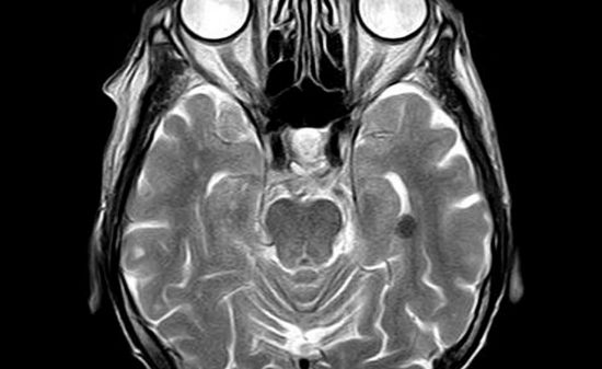
異なるサイズの転移を有する異なる患者について、それぞれ(左から右へ)コントラスト強調3Dグラディエントエコー(CE 3D-GRE)画像、オリジナルBB画像、およびディープラーニングプロセスによって生成されたディープラーニングBB画像を表している。すべての場合において、転移は、オリジナルBB画像およびディープラーニングBB画像の両方でよく観察される。
AIDBの全記事が読み放題のプレミアム会員登録はこちらから↓


(a)オリジナルBB画像の偽陽性(誤検知)の結果
(b)ディープラーニングBB画像の偽陰性(検知漏れ)の結果
症例1では、オリジナルBB画像は、転移に似た左前頭葉において高い信号強度(矢印)を示したが、これは、コントラスト強調3Dグラディエントエコー(CE 3D-GRE)で見られるように、溝の不完全な血管抑制によるものであった。ディープラーニングBB画像は対応する領域に異常を示さなかった。4ヵ月後の追跡調査においても、疑われる転移性病変はまだ認められない。 症例2では、オリジナルのBB画像は左側頭葉に小さな高信号強度の病変(矢印)を示しているが、これはディープラーニングBB画像では見られなかった。追跡調査の前に、隣接する皮質血管のために放射線科医がCE 3D-GRE画像上の病変を識別することができなかった。これは、ディープラーニングBBが病変を見せなかった理由を説明するかもしれない。病変は2ヵ月後に追跡調査で明らかになり、転移として確認された。
著者
Yohan Jun (School of Electrical and Electronic Engineering, Yonsei University, Seoul, Korea)
Taejoon Eo (School of Electrical and Electronic Engineering, Yonsei University, Seoul, Korea)
Taeseong Kim (School of Electrical and Electronic Engineering, Yonsei University, Seoul, Korea)
Hyungseob Shin (School of Electrical and Electronic Engineering, Yonsei University, Seoul, Korea)
Dosik Hwang (School of Electrical and Electronic Engineering, Yonsei University, Seoul, Korea)
So Hi Bae (Department of Radiology, Severance Hospital, Research Institute of Radiological Science and Center for Clinical Image Data Science, Yonsei University College of Medicine, Seoul, Korea)
Yae Won Park (Department of Radiology, Ewha Womans University College of Medicine, Seoul, Korea)
Ho-Joon Lee (Department of Radiology, Severance Hospital, Research Institute of Radiological Science and Center for Clinical Image Data Science, Yonsei University College of Medicine, Seoul, Korea)
Byoung Wook Choi (Department of Radiology, Severance Hospital, Research Institute of Radiological Science and Center for Clinical Image Data Science, Yonsei University College of Medicine, Seoul, Korea)
Sung Soo Ahn (Department of Radiology, Severance Hospital, Research Institute of Radiological Science and Center for Clinical Image Data Science, Yonsei University College of Medicine, Seoul, Korea)
出版情報
Received: 22 November 2017 / Accepted: 5 June 2018 / Published: 21 June 2018
Open Access This article is licensed under a Creative Commons Attribution 4.0 International License, which permits use, sharing, adaptation, distribution and reproduction in any medium or format, as long as you give appropriate credit to the original author(s) and the source, provide a link to the Creative Commons license, and indicate if changes were made. The images or other third party material in this article are included in the article’s Creative Commons license, unless indicated otherwise in a credit line to the material. If material is not included in the article’s Creative Commons license and your intended use is not permitted by statutory regulation or exceeds the permitted use, you will need to obtain permission directly from the copyright holder. To view a copy of this license, visit http://creativecommons.org/licenses/by/4.0/.
■サポートのお願い
AIDBを便利だと思っていただけた方に、任意の金額でサポートしていただけますと幸いです。


 PAGE TOP